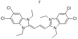
3520-43-2
 3520-43-2 結(jié)構(gòu)式
基本信息
可見光吸收劑515
JC-1(線粒體膜電位探針)
線粒體膜電位熒光探針JC-1
5,5',6,6'-四氯-1,1',3,3'-四乙基苯并咪唑羰花青碘化物
5,5',6,6'-四氯-1,1',3,3'-四乙基苯并咪唑羰花菁碘化物
CBIC2(3),5,5′,6,6′-TETRACHLORO-1,1′,3,3′-TETRAETHYL-IMIDACARBOCYANINE IODIDE
JC-1, >95%
JC-1 jodide
CBIC2, >95%
-tetraethyl-5,5'
-tetrachloroimidacarbocyanine iodide
5,5',6,6'-TETRACHLORO-1,1',3,3'-*TETRAETHYBENZIMIDA
1,10,3,30-tetraethyl-5,50,6,60-tetrachloroimidacarbocyanine Iodide
5,5',6,6'-tetrachloro-1,1',3,3'-tetraethyl-imidacarbocyanine iodide
Imidacarbocyanine Iodide, 1,10,3,30-tetraethyl-5,50,6,60-tetrachloro-
物理化學(xué)性質(zhì)
| 報價日期 | 產(chǎn)品編號 | 產(chǎn)品名稱 | CAS號 | 包裝 | 價格 |
| 2024/11/08 | HY-15534 | JC-1 JC-1 | 3520-43-2 | 1mg | 700元 |
| 2024/11/08 | HY-15534 | JC-1 JC-1 | 3520-43-2 | 2mg | 1200元 |
| 2024/11/08 | HY-15534 | JC-1 JC-1 | 3520-43-2 | 5 mg | 2400元 |
常見問題列表
Guidelines (Following is our recommended protocol. This protocol only provides a guideline, and should be modified according to your specific needs).
Labeling of Cells:
1. Culture cells in 6-, 12- , 24-, or 96-well plates at a density of 5× 10
5
cells/mL. Incubate the cells according to your normal protocol.
2. Ensure that the JC-1 and DMSO has equilibrated to room temperature, and then prepare a 200 μM stock solution by dissolving the contents of one vial in 230 μL of the DMSO provided.
3. For the control tube, allow the vial of CCCP has come to room temperature, add 1 μL of CCCP (50 mM). Incubate cells at 37°C for 5 minutes.
4. Add 10 μL JC-1 (200 μM) per well to make the final concentration at 2 μM. Incubate cells at 37°C, 5% CO
2
, for 15-20 minutes. If additional labeling followed, for example with an annexin V, begin with step 2.a. If not, proceed with step 1.e.
5. After incubation, centrifuge cells for 3-4 minutes at 400× g at 4°C, carefully aspirate the supernant.
6. Wash cells twice with PBS (1×): add 2 mL PBS (1×) to suspend cells and vortex to mix thoroughly. Centrifuge cells for 3-4 minutes at 400× g at 4°C, carefully aspirate the supernant.
7. Add 500 μL PBS (1×) to suspend cells. Analyze sample on a flow cytometer, fluorescence microscopy, or fluorescence microplate reader.

