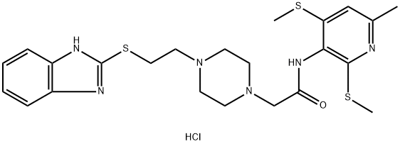
217094-32-1
 217094-32-1 結(jié)構(gòu)式
常見問題列表
|
ACAT-1 0.45 μM (IC 50 ) |
ACAT-2 102.85 μM (IC 50 ) |
The potency and selectivity of K-604 for human ACAT-1 and ACAT-2 is examined. The IC 50 value of K-604 for human ACAT-1 is 0.45 μM and that for human ACAT-2 is 102.85 μM, indicating that K-604 is 229-fold more selective for ACAT-1 than ACAT-2. Kinetic analysis indicates that the inhibition is competitive with respect to oleoyl-coenzyme A with a K i value of 0.378 μM. K-604 efficiently inhibits cholesterol esterification in human macrophages with IC 50 value of 68 nM. In cell free biochemical assays, K604 at 0.5 μM inhibits ACAT1 enzymatic activity by 70% without significantly inhibiting the ACAT2 enzyme activity. To investigate whether blocking ACAT1 increases autophagy in neuronal cells, N2a cells are treated with K604. The result shows that at 0.1 to 1 μM, K604 inhibits ACAT activity by 60-80%. Next K604 is added to N2a cells at concentrations from 0.1 to 1 μM for 24 h, and LC3 levels are examined by western blot. The result shows that K604 increases the LC3-II/LC3-I ratio, a reliable marker for autophagosome formation, in a dose-dependent manner. The number of the fluorescent LC3 puncta is significantly increased in N2a cells after K604 treatment. By western blot, K604 significantly decreases the levels of p62 in N2a cells.
Using F1B hamsters, an animal model susceptible to diet-induced hyperlipidemia and atherosclerosis, the effects of K-604 on aortic lesion areas and plasma cholesterol levels are assessed. Administration of K-604 does not affect body weight or food consumption. The plasma cholesterol levels in fat-fed hamsters are ~12-fold higher than those in chow-fed hamsters, which are significantly decreased by K-604 only at the highest dose tested (30 mg/kg) but not at lower doses (1-10 mg/kg).The fatty streak lesions stain with oil red O are markedly induced by the high-fat diet, which is significantly reduced by administration of K-604. Further, the histological analyses of the atherosclerotic lesions are performed. The fatty streak lesions in the control group are characterized by accumulation of foamy macrophages in the subendothelial space. In contrast, the areas occupied by foamy macrophages are markedly reduced by administration of K-604.
