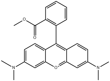
115532-49-5
 115532-49-5 結(jié)構(gòu)式
基本信息
6-(dimethylamino)-9-(2-methoxycarbonylphenyl)xanthen-3-ylidene]-dimethylazanium
常見問題列表
TMRM is a fluorescent probe (excitation, 530±21 nm; emission, 592±22 nm). The fluorescence signal in the presence of safranin or TMRM shows a slight decrease after the addition of glutamate, indicative of increased polarization of the mitochondrial inner membrane. In the presence of TMRM (2 μM) the coupled respiration with Complex I substrates or upon the addition of Complex II substrate is decreased by 27%. Exposure of hippocampal cultures to low concentrations of TMRM (50 to 500 nM) for 1 to 3 hours results in selective staining of mitochondria in both neurons and the underlying glial cells. Exposure of hippocampal cultures to high concentrations of TMRM (1 to 25 μM) stains mitochondria selectively and quickly, reaching a plateau after 5 to 10 min. Low concentrations of TMRM (50 to 200 nM) do not induce apoptosis, whereas higher concentrations (0.5 and 2.5 μM) enhance apoptosis (K D =?500 nM).